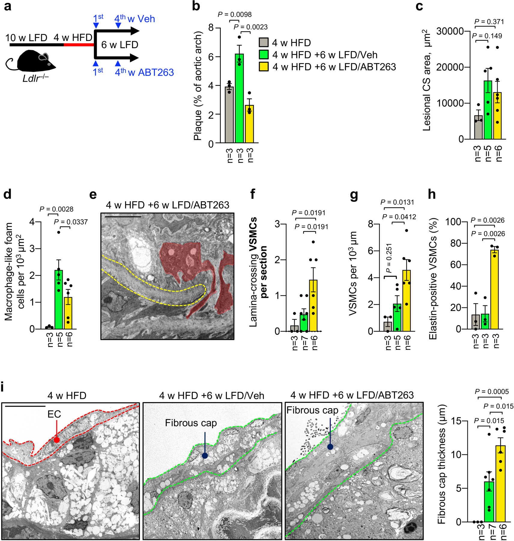
Extended Data Fig. 8: ABT263 accelerates formation of a fibrous cap structure by stimulating migration of medial VSMC.

a, Schematic of experiments designed to study the impact of ABT263-mediated senolysis on fibrous cap formation in early inner aortic arch lesions of Ldlr−/− mice after HFD-to-LFD switching. b, Total plaque burden as measured by en face scoring in mice from indicated groups. c, Neointimal cross sectional area as measured by TEM in lesions of indicated mice (legend is as in b). d, Macrophages per neointimal area in mice of indicated groups (legend is as in b). e, Representative image illustrating VSMCs traversing the first elastic lamina (red masks) in a lesion of the indicated Ldlr−/− mouse (quantified in f). f, Quantification of VSMCs crossing the first elastic lamina in mice of indicated groups (legend is as in b). g, VSMCs per μm of neointima in mice of indicated groups (legend is as in b). h, Percentage of elastin-producing VSMCs in mice from indicated groups (legend is as in b). i, (Left) Representative electron micrographs of 4-week HFD simple foam cell lesions and lesions remodeling during 6-week LFD feeding with Veh or ABT263 administration (quantified in right panel). EC, Endothelial cell. (Right) Quantification of fibrous cap thickness (legend is as in b). “n” refers in all panels to number of mice. All analyses were performed by ordinary one-way ANOVA with Holm-Sidak multiple comparison correction for the indicated comparisons. Error bars represent s.e.m. Scale bars in e and i are 5 μm.
