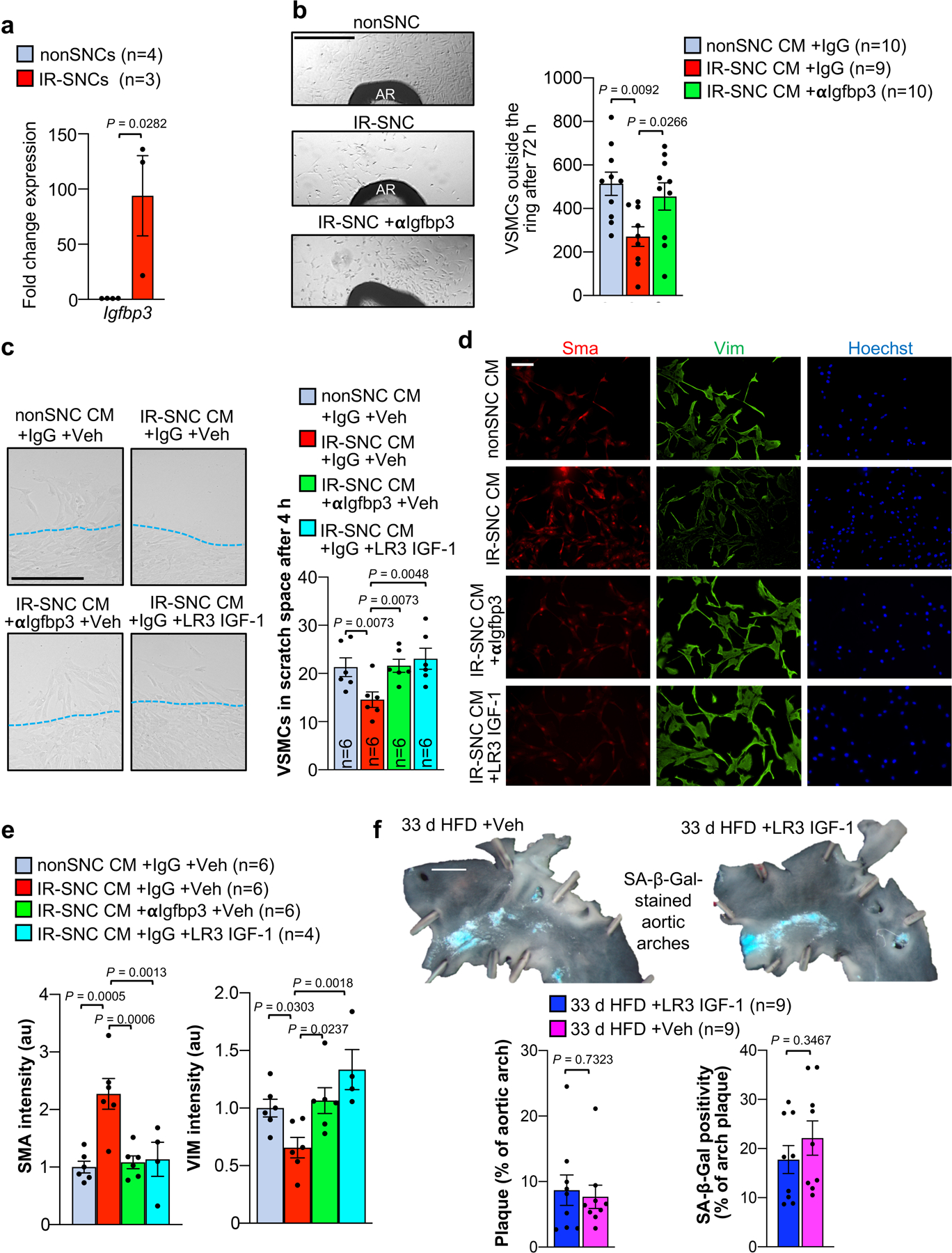
Extended Data Fig. 9: Senescent cell-derived IGFBP3 inhibits IGF1-mediated promigratory phenotype switching of VSMCs.

a, Expression of Igfbp3 in the indicated MEFs. b, (Left) Representative images of VSMC outgrowing from aortic rings (AR) of wildtype C57BL/6 mice treated with the indicated conditioned media (CM). (Right) Quantification of outgrowing VSMCs in the indicated treatment groups. c, (Left) Representative images of human aortic VSMCs emigrating into scratch wound space with indicated conditioned media (CM). Red dashed lines indicate cell monolayer/scratch wound boundary. (Right) Quantification of emigrating VSMCs in the indicated experimental groups. LR3 IGF-1, long R3 mutant stabilized recombinant IGF-1. d, Immunofluorescent staining of Vim and Sma in human aortic VSMC with the indicated treatments. CM, conditioned media (quantified in e). e, Quantification of average fluorescent signal intensity of SMA and VIM per cell in human aortic VSMC receiving indicated treatments. f, (Top) Representative images of SA β-Gal stained Ldlr–/– aortic arches from mice fed HFD for 33 d with concurrent administration of either LR3 IGF1 or vehicle control. (Bottom left) Quantification of total plaque burden in the aortic arch and (bottom right) percentage of aortic arch plaque with SA β-Gal positivity in indicated treatment groups. “n” refers individual MEF lines (panel a); individual aortic rings in b; individual mice in panel f; and, individual lines of MEF conditioned media (panels c and e). Panel a was analysed with unpaired, two-tailed t-test. Panel f was analyzed by unpaired, two-tailed t-test with Welch’s correction. Analyses in panels b and e were performed by ordinary one-way ANOVA with Holm-Sidak multiple comparison correction for the indicated comparisons, and analysis of panel c was performed by RM one-way ANOVA with Holm-Sidak multiple comparison correction for the indicated comparisons. Error bars represent s.e.m. Scale bar in b is 500 μm; c, 200 μm; d, 100 μm; and, f, 1 mm.
