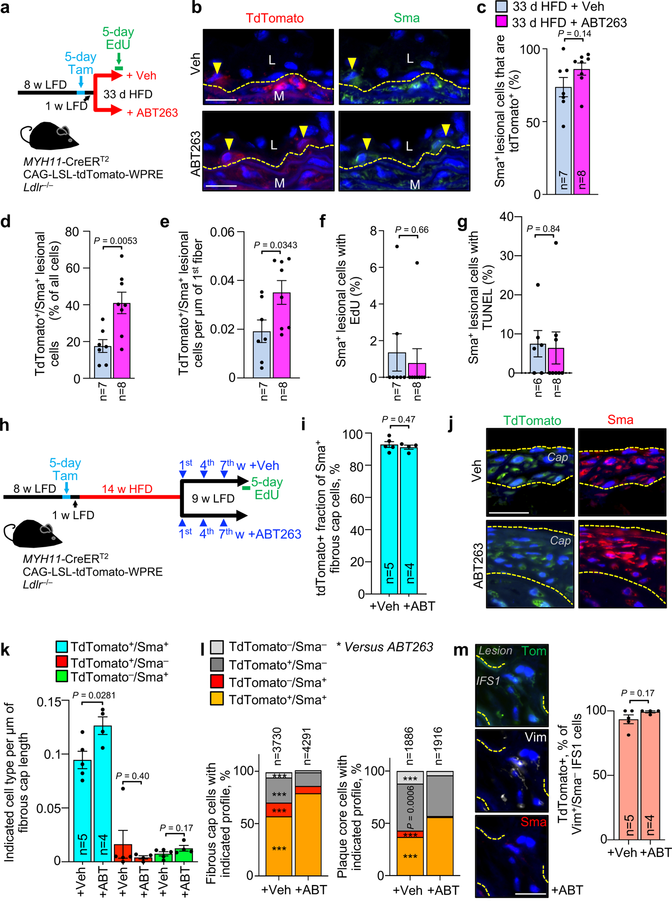
Figure 5: VSMC lineage tracing approaches reveal senolysis enhances medial VSMC phenotypic switching and migration into lesions.

a, Schematic of the experimental design for data in b-g. b, Representative images of inner aortic arch lesions of the indicated mice (quantified in c) immunolabeled for tdTomato and Sma. L, lesion; M, media. Arrowheads mark tdTomato+/Sma+ cells; interrupted line mark first elastic fiber. c, Quantification of the percentage of Sma+ neointimal cells in inner aortic arch lesions of mice indicated in a that are also tdTomato+. d, Quantification of the percentage of neointimal cells that are tdTomato+/Sma+ in inner aortic arch lesions of mice indicated in a. e, Quantification of tdTomato+/Sma+ neointimal cells in lesions of mice indicated in a normalized to plaque burdened length of 1st elastic fiber. f, Percentage of neointimal Sma+ cells in mice from a incorporating EdU. g, Percentage of neointimal Sma+ cells in mice from a showing TUNEL+ nuclei. h, Schematic of the experimental design for data in i-m. i, Quantification of tdTomato+ percentage of Sma+ fibrous cap cells of mice from indicated groups. j, Representative images of tdTomato+/Sma+ cells in fibrous caps (bracketed in yellow dashed lines; quantified in k) from indicated groups. k, Quantification of frequency of cells of indicated tdTomato and Sma status per length of fibrous cap for indicated groups. l, Proportion of cells of indicated Sma and tdTomato status in fibrous cap (left) and plaque core (right) from inner aortic arch plaques of mice indicated in h. ***, p <0.001. m, (Left) Representative images of an IFS1 Vim+/Sma−/tdTomato+ cell and (Right) Quantification of the percentage of IFS1 Vim+/Sma− cells that are tdTomato+ for the indicated groups. All panels were analyzed by unpaired, two-tailed t-test with Welch’s correction, but panel l, which was analyzed by global x2, followed up by individual two-tailed Fischer’s exact tests for each combination of Sma and Tom status cells. Error bars represent s.e.m. *, p <0.05; **, p <0.01; ***, p <0.001. “n” always represents number of mice, excepting panel l, where “n” is total cell count. Scale bars in b, j, and m are all 20 μm.
