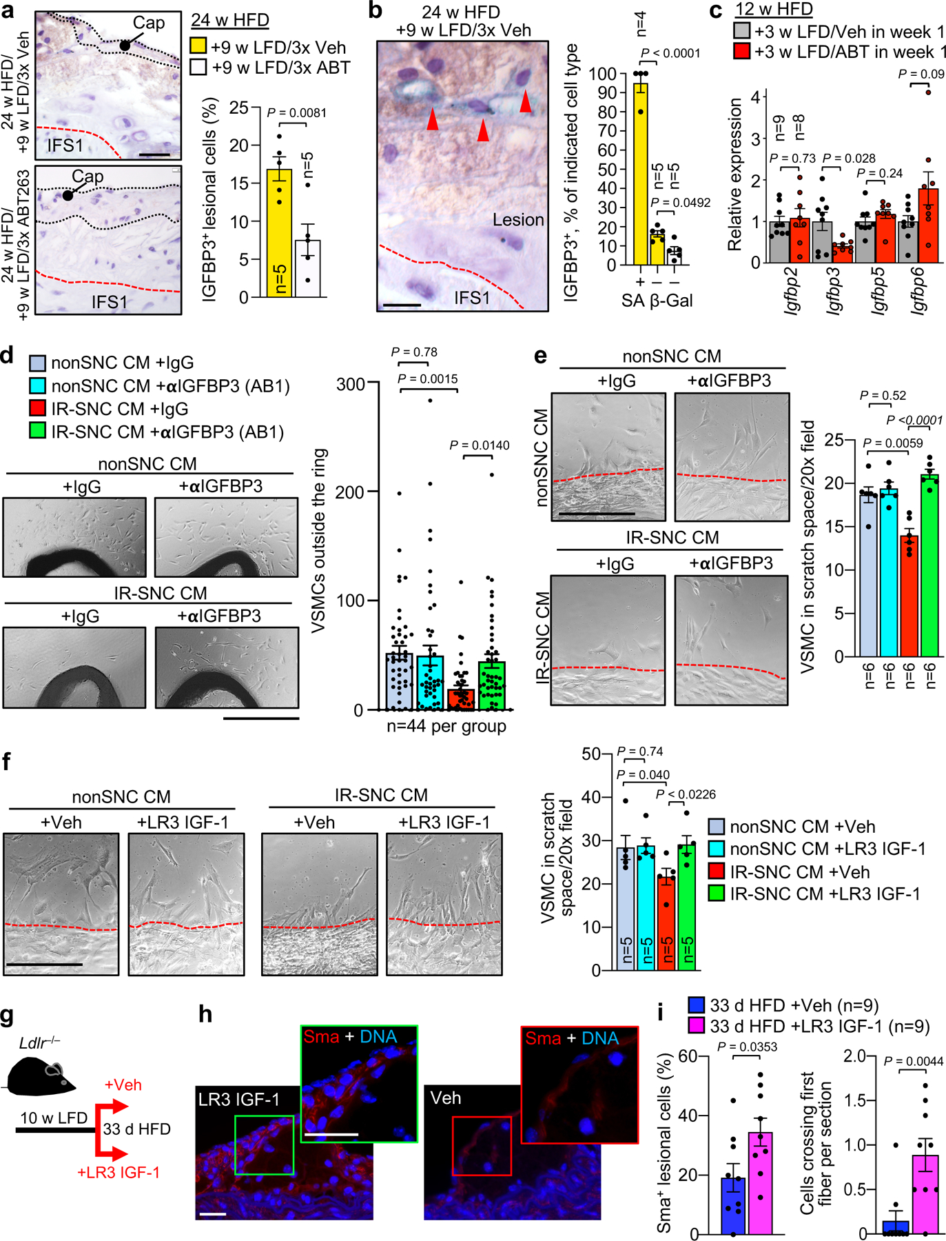
Figure 7: Senolysis depletes lesional Igfbp3 to promote promigratory VSMC phenotype switching of medial VSMCs.

a, (Left) Representative images of lesions of the indicated mice immunostained for Igfbp3. (Right) Quantification of percentage Igfbp3+ cells in lesions of the indicated mice. b, (Left) Representative images of Igfbp3+/SA β-Gal+ lesional cells (arrowheads). (Right) Quantification of Igfbp3+ cell frequency among SA β-Gal+ and SA β-Gal− cells from indicated groups (legend as in a). c, Relative expression of Igfbp genes in brachiocephalic arteries of the indicated Ldlr−/− mice. d, (Left) Representative images of VSMCs outgrowing from aortic rings (AR) of wildtype C57BL/6 mice treated with the indicated conditioned media (CM). αIGFBP3 (AB1), ab193910. (Right) Quantification of outgrowing VSMCs in the indicated treatment groups. e, (Left) Representative images of human aortic VSMCs emigrating into scratch wound space with indicated CM. (Right) Quantification of emigrating VSMCs in the indicated experimental groups (legend as in d). f, (Left) Representative images of human aortic VSMCs emigrating into scratch wound space with indicated CM. (Right) Quantification of emigrating VSMCs in the indicated experimental groups. LR3 IGF-1. g, Schematic of experiments presented in h and i. h, Representative Sma immunostaining in the indicated early lesions. i, (Left) Quantification of neointimal Sma+ cells in lesions of the indicated mice. (Right) Quantification of cells crossing the first elastic fiber in indicated groups. Statistics in panels b and d were performed by ordinary one-way ANOVA with Holm-Sidak multiple comparison correction for the indicated comparisons. Statistics in panels e and f were performed by RM one-way ANOVA with Holm-Sidak multiple comparison correction for the indicated comparisons. All other panels were analyzed by unpaired, two-tailed t-test with Welch’s correction. Error bars represent s.e.m. “n” refers to individual mice in a, b, c, and i; individual aortic rings in d; and, CM from independent MEF lines in e and f. Scale bar in a is 20 μm; b, 10 μm; d, 500 μm; e and f, 200 μm; and, g (main and inset), 20 μm.
