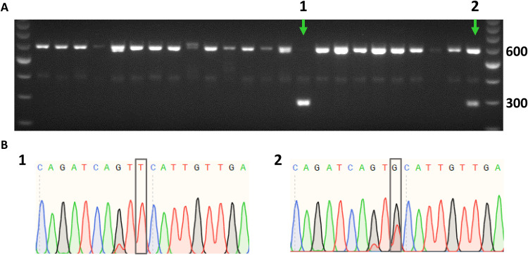
Fig. 2. Introducing identified FN1 mutation in hiPSCs.
Clonal screen for FN1 mutation in hiPSCs by genomic polymerase chain reaction (PCR) followed by Hinc II digestion (A), confirmed by Sanger sequencing (B). The PCR product is 600 base pairs (bp) in size. Upon integration of the provided ssODN, Hinc II digestion results in 303- and 297-bp products. (A) Representative gel image, where FN1 mutant clones are indicated by a green arrow, showing a homozygous clone (lane 1) and a heterozygous clone (lane 2). (B) Sanger sequencing results of homozygous (1) and heterozygous (2) clones, respectively. Gray boxes represent FN1 mutation.

