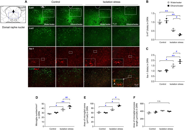
Fig. 3. Serotonergic signals and microglial activation.
After IS with or without ethanol exposure for 28 days, mice were euthanized by transcardial perfusion. Serotonergic and microglial activity was assessed by immunofluorescence analysis of 5-HT and Iba-1 (A) in the dorsal raphe nuclei (DRN), and their intensities were semiquantified (B and C). Microglial cell numbers (D), soma area (E), and length (F) were quantified. The data are expressed as the means ± SD (n = 3). *P < 0.05 and **P < 0.01 compared to the unstressed mice, and #P < 0.05 and ##P < 0.01 compared to the mice not exposed to ethanol.

