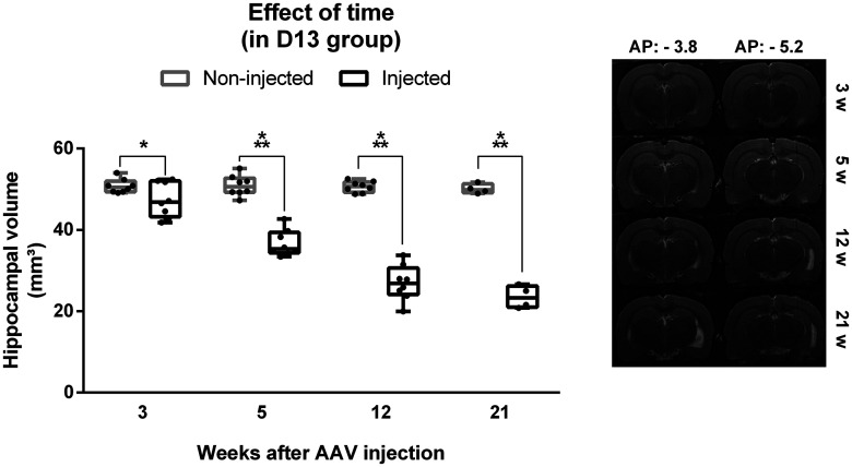
Figure 3.
Effect of time on hippocampal volume in the DREADD group with titer E + 13 (D13). Left, Volume of the injected hippocampus is significantly reduced compared with the non-injected hippocampus at all time points and the difference in hippocampal volume between the injected and non-injected hippocampus increases with time. Dots represent individual animals. Right, Representative example of T2 MRI scans at the anterior and posterior injection sites at different timepoints in one animal. The injected hippocampus is situated in the MR images on the right side; *p < 0.05, ***p < 0.001. AP = anteroposterior.

