Figure 2.

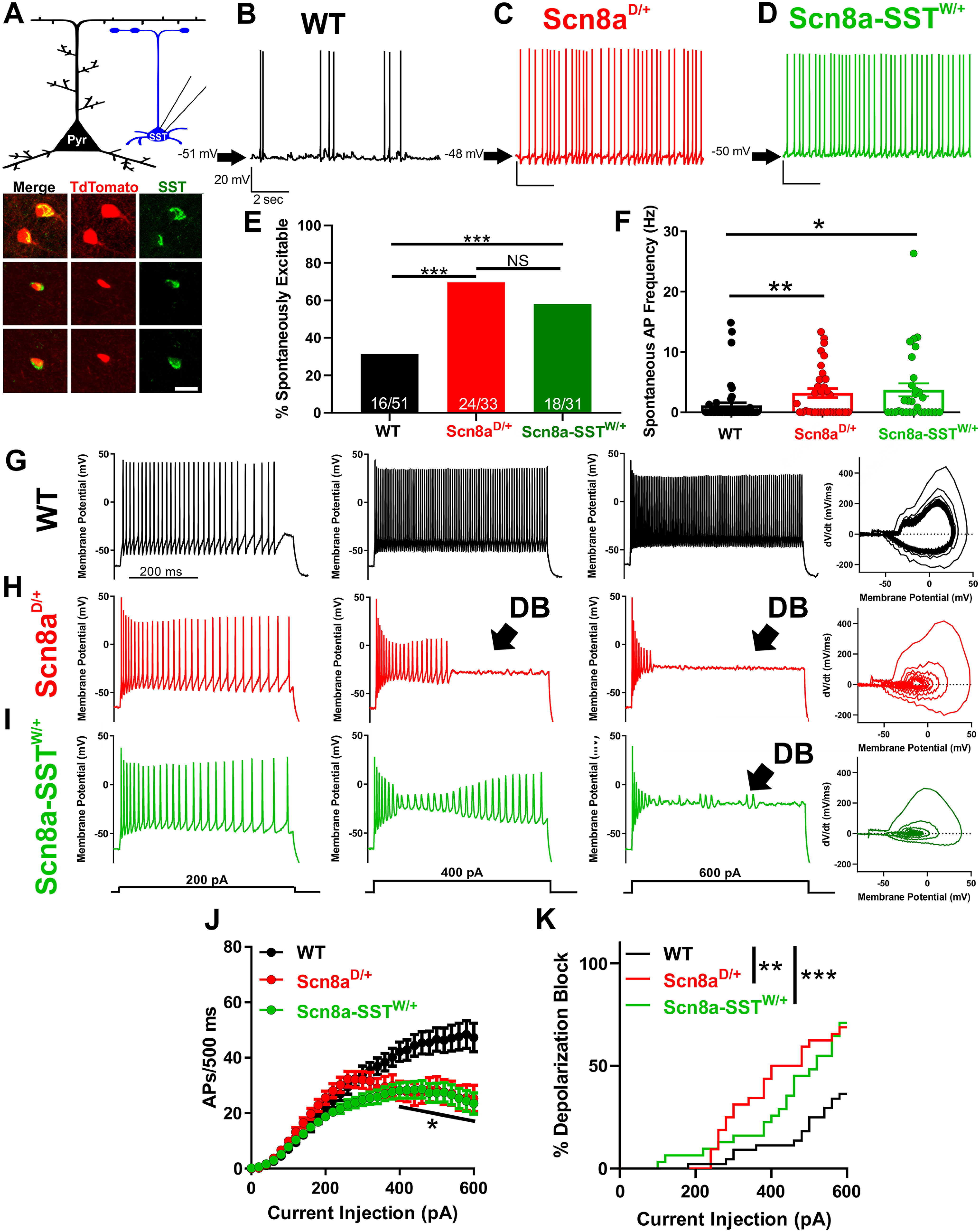
Scn8aD/+ and Scn8a-SSTW/+ SST interneurons are hyperexcitable and readily enter depolarization block. A, Whole-cell recordings collected from WT, Scn8aD/+, and Scn8a-SSTW/+ somatosensory layer V SST interneurons (blue). Example immunohistochemistry images showing colocalization of TdTomato (red) and SST (green) immunofluorescence. Scale bar, 20 μm. B-D, Representative example traces of spontaneous excitability of WT (B; black), Scn8aD/+ (C; red) and Scn8a-SSTW/+ (D; green) SST interneurons. Arrows indicate membrane potential between spontaneous APs. E, Only 16 of 51 (∼31%) WT SST interneurons were spontaneously excitable, whereas 24 of 33 (∼73%) Scn8aD/+ and 18 of 31 (∼58%) Scn8a-SSTW/+ spontaneously fired APs (***p < 0.01 by Fisher's exact test). F, Average spontaneous firing frequencies for WT (black), Scn8aD/+ (red), and Scn8a-SSTW/+ (green) SST interneurons. *p < 0.05, **p < 0.01, Kruskal–Wallis test followed by Dunn's multiple comparisons. G-I, Representative traces for WT (G; black), Scn8aD/+ (H; red), and Scn8a-SSTW/+ (I; green) SST interneurons eliciting APs in response to 500 ms current injections of 200, 400, and 600 pA. Depolarization block is indicated (arrow; DB) in the Scn8aD/+ (H; red) and Scn8a-SSTW/+ (I; green) interneurons. Right, Phase plot corresponding to the 600 pA current traces for WT (G; black), Scn8aD/+ red (H; red), and Scn8a-SSTW/+ (I; green). J, Average number of APs elicited relative to current injection magnitude for WT (black; n = 51, 10 mice), Scn8aD/+ (red; n = 33, 6 mice), and Scn8a-SSTW/+ (green; n = 31, 4 mice). At current injections >400 pA, both Scn8aD/+ and Scn8a-SSTW/+ AP frequencies were reduced relative to WT. *p < 0.05, two-way ANOVA followed by Tukey's correction for multiple comparisons. K, Cumulative distribution of SST interneuron entry into depolarization block relative to current injection magnitude for WT (black), Scn8aD/+ (red), and Scn8a-SSTW/+ (green) mice. Curve comparison by log-rank Mantel-Cox test (**p < 0.01).
