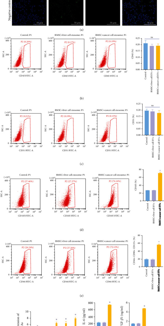
Figure 3.

EVs promoted the differentiation of BMSCs into cancer stem cells. EVs derived from tumor or normal tissues were cocultured with BMSCs for 48 h. Then, (a) the EV uptake was observed by IF staining. Scale bar: 100 μm. 100x magnification. (b) Flow cytometry was performed to test the ratio of CD44 in BMSCs. (c) Flow cytometry assessed the ratio of CD31 in BMSCs. (d) Flow cytometry tested the ratio of CD105 in BMSCs. (e) Flow cytometry assessed the ratio of CD44+ CD90+ CD133+ cells in BMSCs. (f) RT-qPCR evaluated Oct4, SRY, IL-1α, IL-6, and TGF-β1 levels in BMSCs. (g) ELISA was performed to test the content of IL-6 and TGF-β1 in supernatants of BMSCs. ∗P < 0.05 compared to the control. ns indicated no significant change.
