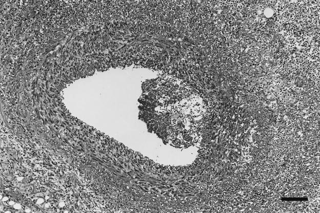
FIG. 2.
Coronary artery of dog 2, infected with B. vinsonii. Shown is the coronary artery with an adherent mass of inflammatory exudate bulging into the arterial lumen, with transmural inflammation of the arterial wall beneath the inflammatory exudate. Severe inflammation surrounding inflamed artery effaces the normal tissue architecture. Hematoxylin and eosin stain; bar = 107 μm.

