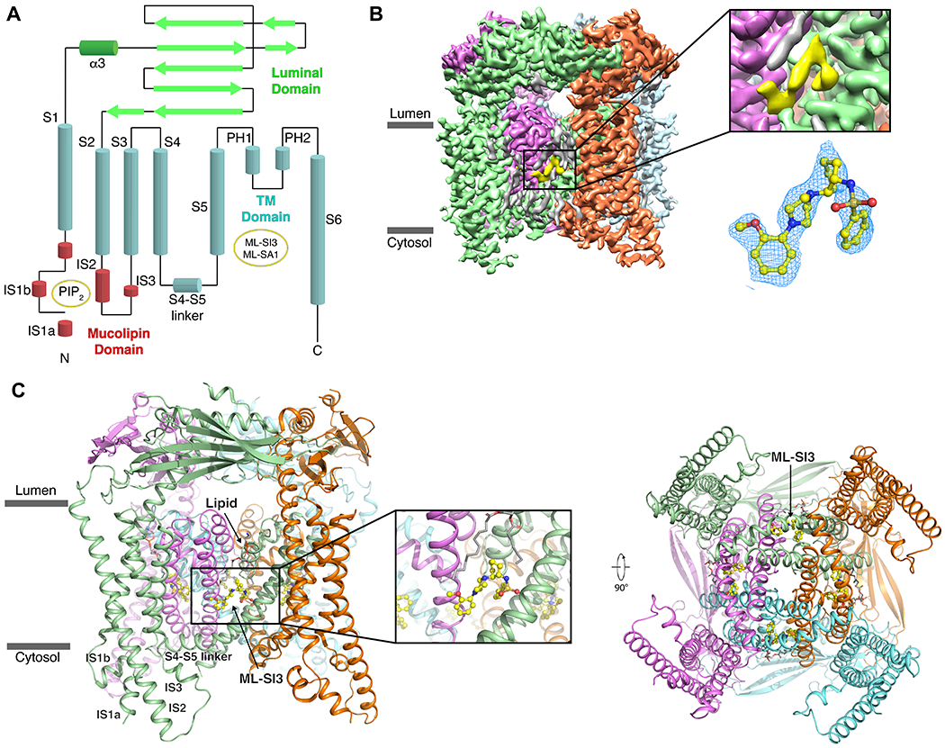
Figure 1. Overall Structure of ML-SI3-bound TRPML1.

(A) A 2D schematic of TRPML1 with the various domains colored, and the binding sites indicated in yellow circles. (B) The cryo-EM map of TRPML1-ML-SI3 with the protomers colored individually and ML-SI3 in yellow. A zoom-in on the density that represents ML-SI3 with the EM-density in mesh (blue) fitted to the molecular structure (yellow sticks). (C) Structural model of TRPML1–ML-SI3 with a zoom-in on ML-SI3 and its binding pocket. Subunits are represented by the same colors as in B.
