
Figure 3.
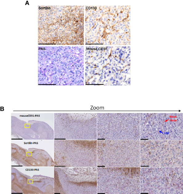
SORT1 and CD133 expression in ES-2 ovarian tumor xenografts. Tumor xenografts were established by subcutaneous inoculation of 7x10⁶ ES-2 ovarian cancer cells, resuspended in 100 μL of HBSS and injected in the right flank of CD-1 nude mice. Tumors were collected when they reached 1500 mm3. (A) Sortilin, CD133, CD31 and PAS stainings were performed as described in the Methods section. (B) Co-staining with PAS was also performed for CD31 (CD31-PAS), sortilin (Sortilin-PAS) and CD133 (CD133-PAS). VM structures (blue arrows) and blood vessels (red arrows) are indicated. Scale bars represent 2500, 250, 100 and 50 µm respectively (left to right column). Representative images are shown from two different ES-2 xenograft tumors.