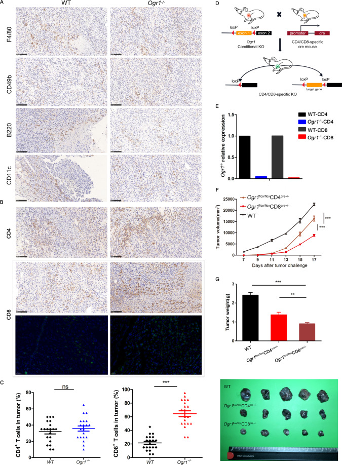
Fig. 3. Ogr1 deletion enhances the infiltration of CD8+ T cells and contributes to tumor regression.
A Representative IHC staining was applied to assess the expression of NK cells (CD49b), macrophages (F4/80), B cells (B220) as well as dendritic cells (CD11c) in xenograft melanomas. B The representative immunohistochemistry and immunofluorescence staining were applied to assess the number of CD4+ T cells and CD8+ T cells infiltration in xenograft melanomas. C Staining intensities for CD4+ T cells and CD8+ T cells in melanomas were determined by the assignment of semiquantitative scores. D Schematic of mouse models of conditional knockout of Ogr1 in CD4+ and CD8+ T cells. When Ogr1 flox mice mated with CRE mice, the specific exon of Ogr1 was deleted, thus realizing the conditional knockout of Ogr1 gene. E Ogr1 expression in T cells was detected by RT-PCR. Tumor growth (F) and mean tumor volume (Day 20) (G) of subcutaneous B16-F10 (5 × 10⁵ in 100 µl PBS) in WT mice versus Ogr1flox/floxCD4Cre+/− or Ogr1flox/floxCD8Cre+/− mice. In A and B, original magnification ×20; scale bar = 100 μm. F, G experiments were repeated three times. The results are presented as means ± SD. *P < 0.05, **P < 0.01, ***P < 0.001, as obtained by unpaired test.

