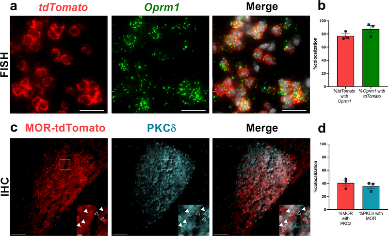
Figure 4 – A large subset of MOR lineage neurons (Opmr1-tdTomato) in the CeA express PKCδ.
(a) Dual FISH of Oprm1 (green) and tdTomato (red) mRNA, plus DAPI counterstaining (white) in the CeA, revealed that a majority of tdTomato+ neurons express Oprm1 mRNA (scale bar = 25 μm). (b) Quantification of a (n = 3 mice, 8–10 sections per mouse). (c) Representative images demonstrating MOR-tdTomato and PKCδ immunofluorescence in the CeA. Insets are cropped, enlarged images of the boxed region. Closed arrows denote MOR-tdTomato/PKCδ colocalization, and open arrows denote MOR-tdTomato in the absence of PKCδ. Scale bars = 100 μm; inset = 10 μm. (d) Quantification of percentage colocalization of MOR-tdTomato and PKCδ (n = 3 mice, 6 to 8 sections per mouse).

