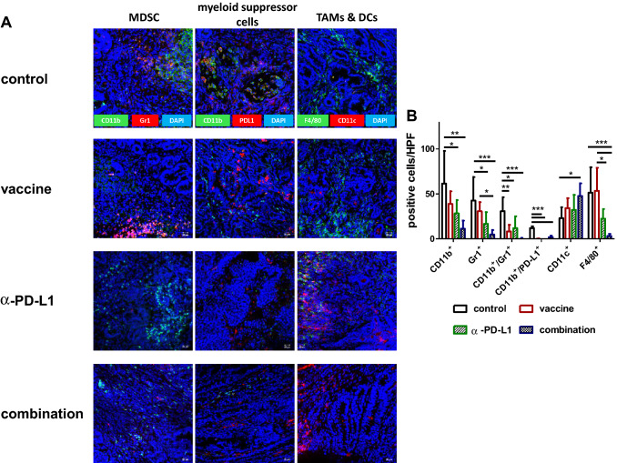
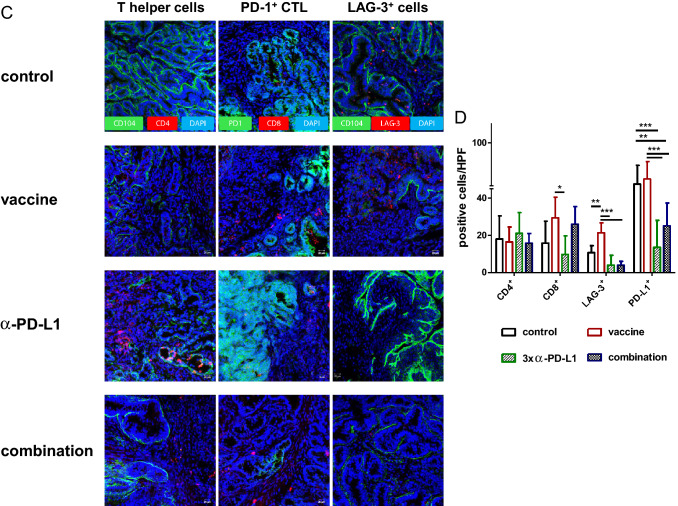
Fig. 6.
Immunofluorescence a, c Residual Mlh1−/− GIT were resected after therapeutic vaccination and cryostat sections of 4 µm prepared. Tumor microenvironment was studied upon staining with specific mAbs, followed by nuclear staining with DAPI. Pictures were taken on a laser scanning microscope (Zeiss) using 20× objectives. b, d Quantification of infiltrating immune cells. At least three pictures were
taken from each slide and numbers of infiltrating cells counted. Data are given as infiltrates/HPF. Mean + SD, n ≥ 3 samples/group; *p < 0.05, **p<0.01; ***p<0.001 one-way ANOVA (Bonferroni’s multiple comparison test)


