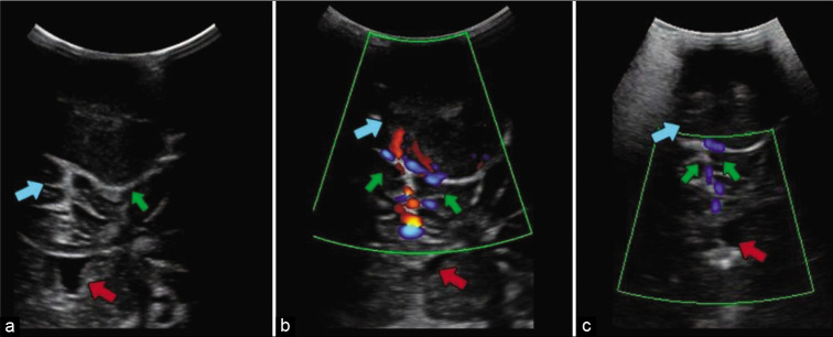
Figure 2:
Ultrasound image (a) showing the exophytic lesion (green arrow) in direct contact with the cerebral parenchyma (blue arrow). Bone erosion created a window in the frontal bone through which even the lateral ventricles (red arrow) are visible. Doppler ultrasound performed before embolization (b) showing vascularity of the tumor (green arrows) and after embolization (c) showing reduced blood flow (green arrows). In these images, the lesion (green arrow and blue arrows, a and b image respectively) and the lateral ventricles (red arrows) surrounded by brain parenchyma are still seen.

