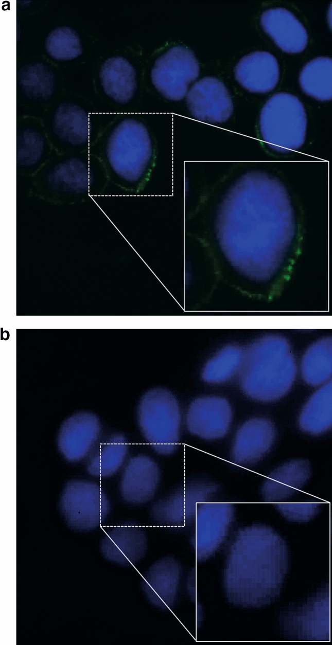
Figure 2.

Spike-adhesion to the Caco-2 cell surface in the absence (a) or presence (b) of diosmectite, as shown by immunofluorescence microscopy. Spike protein RBD is shown after being probed with the primary and secondary fluorescein isothiocyanate-conjugated antibody (green). Nuclei were stained using Hoechst (blue). Images shown are × 1000 magnification. A detail of a single cell is shown in the square. (a) Caco-2 cells incubated with spike protein RBD. (b) Caco-2 cells pre-incubated with diosmectite and co-incubated with spike protein RBD.
