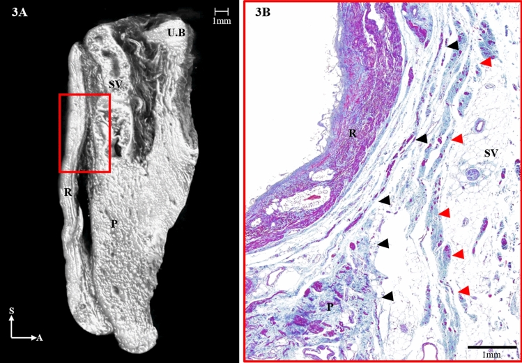
Figure 3.
(A) Three-dimensional anatomy of the Denonvilliers’ fascia (DVF; red) from a sagittal section reconstructed from micro-computed tomography. (B) The distal, seminal part of the DVF is shown diverging to the anterior sheet and merging with the prostatic capsule (red arrowheads) and the posterior sheet (black arrowheads), covering the rectum. UB, urinary bladder; SV, seminal vesicle; P, prostate; R, rectum; S, sagittal; A, anterior.

