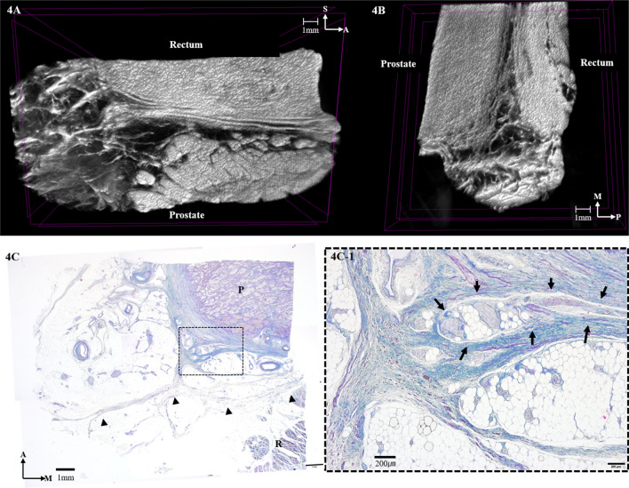
Figure 4.
Three-dimensional anatomy of the Denonvilliers’ fascia (DVF) at the level of the prostate reconstructed from micro-computed tomography. (A) Horizontal section. (B) Sagittal section. (C) Masson trichrome-stained section at the level of the prostate. Horizontal sections of the DVF are indicated by the black arrowheads. Detail window C-1 shows that DVF is as closely related to the prostatic capsule and the neurovascular bundles, which are indicated by the black arrows. P, prostate; R, rectum; M, medial; A, anterior; P, posterior.

