Fig. 2.
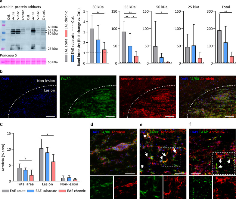
Acrolein-protein adduct formation coincides with high levels of inflammation and microglia/macrophage accumulation in different stages of EAE. a Detection of acrolein-protein adducts by western blot in different stages of EAE compared to healthy controls (dashed line). n = 7–8 animals/group. Acrolein-protein adducts were increased in EAE vs. healthy controls (statistics not shown), except for subacute and chronic EAE at 60 kDa, and chronic EAE at 50 kDa. b Immunohistochemistry demonstrating acrolein-protein adducts in hypercellular EAE spinal cord lesions characterized by an abundance of F4/80+ microglia/macrophages. c Quantification of acrolein-protein adduct immunoreactivity (% positive vs. total area) in EAE spinal cord sections, showing higher acrolein-protein levels in lesion vs. non-lesion areas, and in acute vs. chronic EAE. n = 6 animals/group. d Acrolein-protein adducts in perivascular cuff containing F4/80+ microglia/macrophages. e Acrolein-protein adducts in brain parenchyma co-localized with F4/80+ microglia/macrophages (arrowheads) and with astrocytic processes that appeared F4/80− (arrows), but GFAP+ (arrows in f). Data are mean ± SD. One-way ANOVA or Kruskal–Wallis, post hoc testing *p < 0.05, **p < 0.01 between the indicated groups. Scale bars are 100 µm (b) or 50 µm (d–f)

