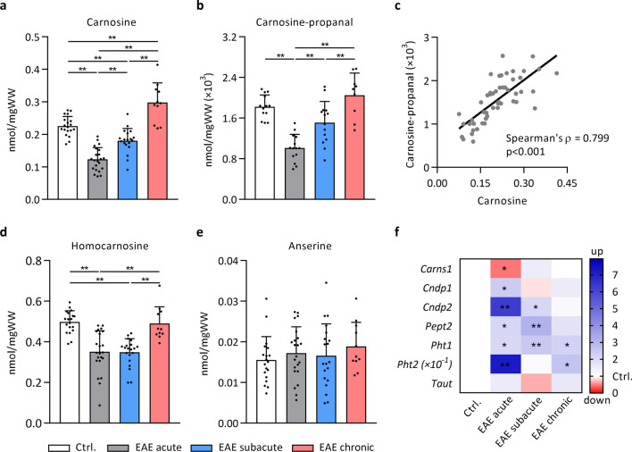
Fig. 3.
Spinal cord carnosine and carnosine-propanal are depleted in acute EAE. UPLC-ESI–MS/MS-based quantification of histidine-containing dipeptides and their carbonyl conjugates shows changes in a, c carnosine, b, c carnosine-propanal and d homocarnosine, but not e anserine, in different stages of EAE compared to healthy control mice. n = 8–23 animals/group. Carnosine-propanal values (~ 0.0015 nmol/mgWW) were transformed (× 103) to facilitate visualisation on the Y axis. f mRNA expression of key regulators involved in carnosine homeostasis was altered in EAE spinal cord compared to healthy controls. Gene expression data are fold changes vs. control mice. Due to high expression of Pht2 in EAE mice, data were transformed (× 10–1) to allow visualisation. n = 6–7 animals/group. Carns1, carnosine synthase; Cndp1, carnosine dipeptidase 1; Cndp2, cytosolic non-specific dipeptidase 2; Pept2, peptide transporter 2 (Slc15a2); Pht1, peptide/histidine transporter 1 (Slc15a4); Pht2, peptide/histidine transporter 2 (Slc15a3); Taut, taurine transporter (Slc6a6). Data are mean ± SD. One-way ANOVA or Kruskal–Wallis, post hoc testing *p < 0.05, **p < 0.01 between the indicated groups (a, b, d, e) or vs. healthy control mice (f)

