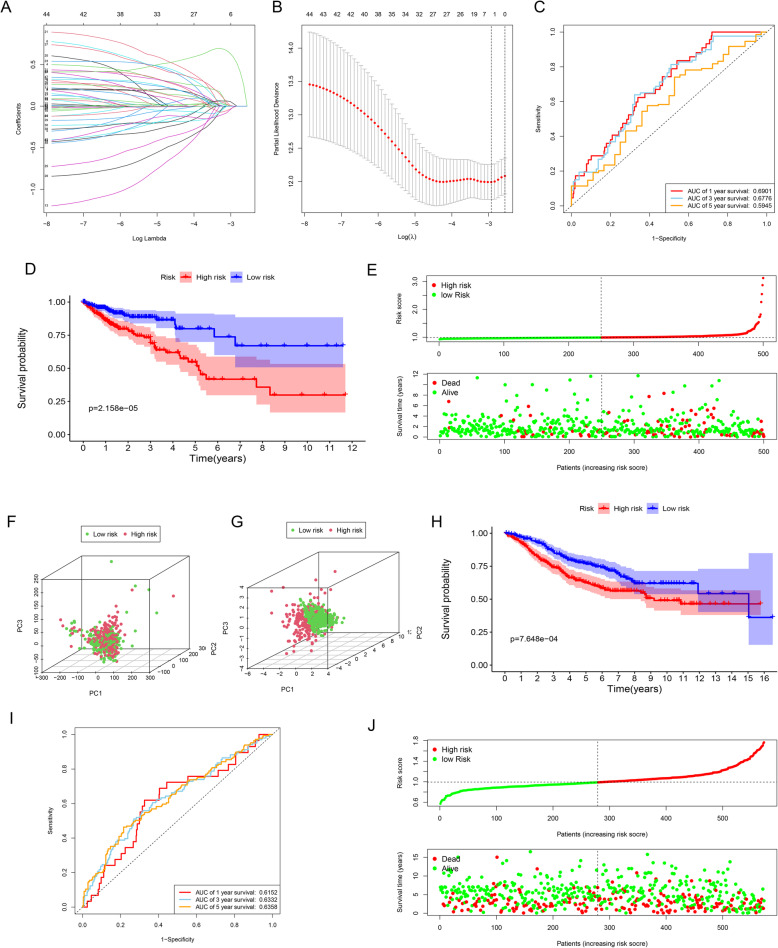
Fig. 3.
Development and validation of the risk signature. (A) LASSO Cox regression coefficient profiles of the 47 LMRG. Each curve represents the changing trajectory of one LMRG. (B) 10-fold cross-validation for tuning parameter selection in LASSO model. Each red dot represents a lambda value with a confidence interval. The two dotted lines indicate the optimal values by minimum criteria and 1-SE criteria by 10-fold cross-validation (standard error; SE). The Y-axis shows partial likelihood deviance values with error bars, and the X-axis shows the penalization coefficient (logλ). (C) Time-dependent ROC curves for 1-, 3-, and 5-year OS in the training group. (D) The survival curves showed significant differences between the high- and low-risk groups (P < 0.001). (E) Risk score distribution (above) and survival status (below) of CRC patients in different risk groups. (F) PCA showed no significant difference in risk status of CRC patients on the basis of the whole gene set. Different color points represent patients with different risk groups. (G) PCA showed that the high-risk group could be distinguished effectively from the low-risk groups based on the risk signature. (H) The survival curves showed significant differences between high- and low-risk patients in the validation set (P < 0.001). (I) Time-dependent ROC curves for 1-, 3-, and 5-year OS in the validation set. (J) Risk score distribution (above) and survival status (below) of CRC patients in the validation set

