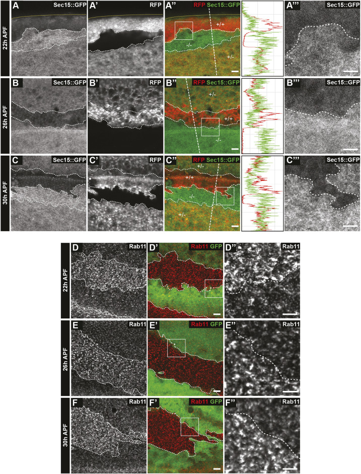
Fig. 3.
Accumulation of Sec15 and Rab11 in RASSF8 mutant pupal wing clones. (A-C‴) Increase in Sec15::GFP (driven by the ubiquitin promoter) in RASSF8 mutant clones (negative for RFP in red) at 22 (A-A″), 26 (B-B″) and 30 (C-C″) h APF. Clone boundaries are marked by white dotted lines. Yellow dotted lines at 22 h AFP show the edge of the wing. In the merge channel, the genotypes of the clones are given [+/+, wild type (two copies of RFP); +/−, heterozygous (one copy of RFP); −/−, homozygous RASSF8 mutant (no copies of RFP)]. A‴, B‴ and C‴ are zoomed-in views of the boxed areas in A″, B″ and C″, respectively. Traces show the intensity profiles at the straight white dashed lines in the merged images using Fiji. (D-F″) Accumulation of Rab11 in RASSF8 mutant clones. Rab11 antibody staining in RASSF8 mutant clones marked by the absence of GFP at 22 (D-D″), 26 (E-E″) and 30 (F-F″) h APF. D″, E″ and F″ are zoomed-in views of the boxed areas in D′,E and F′, respectively. Scale bars: 10 μm in A,A″,B,B″,C,C″,D,D′,E,E′,F,F′; 7 μm in A‴,B‴,C‴,D″,E″,F″.

