Fig. 6.
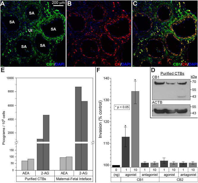
Expression of cannabinoid ligands and receptors at the maternal-fetal interface and the functional consequences. (A-C) Tissue sections were immunostained for CNR1 (CB1) and cytokeratin (CK), a trophoblast marker. Nuclei were localized with DAPI. Each panel was a confocal z-stack maximum intensity projection. Anti-CNR1 specifically reacted with invasive/endovascular CTBs in the walls of uterine (Ut) spiral arteries (SA). Cytotrophoblasts (CTBs) in the cell columns showed variable CNR1 expression, while syncytiotrophoblasts were negative (data not shown). The results shown were representative of the immunostaining observed in a minimum of three biological replicates. (D) Immunoblotting with the same antibody detected a band of the expected molecular weight in CTB lysates. Protein loading was estimated by immunoblotting with anti-actin-β (ACTB). Cells from three placentas were analyzed. (E) Mass spectrometry enabled measuring the absolute amounts of the major mammalian endogenous cannabinoids, anandamide (AEA) and 2-arachidonoylglycerol (2-AG), in isolated CTBs and biopsies of the maternal-fetal interface that included invasive CTBs. The results from replicate samples are shown. (F) Among the CNR1 and CNR2 agonists and antagonists assayed, only a CNR1 agonist had a dose-dependent effect increasing invasion (n=3 biological replicates, *P<0.05, unpaired, two-tailed Student's t-test).

