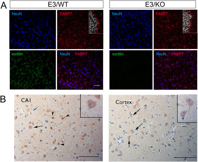
Fig. 5.
Expression of FABP7 in neurons of mouse and human brains. (A) Immunohistological detection of FABP7 (red) in brain cortical sections from apoE3 mice, either wild-type (WT) or genetically deficient for Sort1 (KO). Additionally, the sections were stained for the neuronal marker NeuN (blue) and sortilin (green) as well as DAPI (white; in insets). Merged images show co-expression of FABP7 with NeuN. As a positive control, the insets document expression of FABP7 in glia in the cerebellum of E3/WT and E3/KO mice. Representative images from analysis of three mice per genotype are shown. Scale bar: 100 µm. (B) Staining for FABP7 in hippocampal subfield CA1 and temporal cortex specimens of AD patients, showing immunoreactivity in glial cells (arrowheads) and light positivity in sparse neuronal cells (arrows, and insets). Representative images from one of three AD cases analyzed are shown (pathological characteristics given in Table S3). Scale bars: 100 µm.

