Fig. 3.
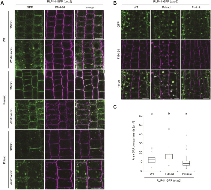
RLP44–GFP variants undergo endocytosis. (A) Fluorescence derived from RLP44–GFP WT, Pdead and Pmimic variants accumulates in enlarged structures after 20 µM wortmannin (Wm) treatment for 165 min, suggesting they reach late endosomes. Note increased plasma membrane labelling of RLP44–GFP Pdead after Wm treatment. Scale bars: 10 µm. (B) Fluorescence derived from RLP44–GFP WT, Pdead and Pmimic variants accumulates in BFA bodies. Roots were treated with 50 µM of BFA or DMSO for 120 min and with FM4-64 for 20 min before imaging. Scale bars: 10 µm. (C) Image quantification reveals largest fluorescent area in BFA bodies of RLP44–GFP-derived fluorescence (lower panel), n=116 (WT), n=102 (pdead), n=90 (Pmimic) measurements in 18 independent roots for each genotype. Boxes indicate range from 25th to 75th percentile, horizontal line indicates the median, whiskers indicate data points within 1.5 times the interquartile range. Markers above whiskers indicate outliers. Different letters indicate statistically significant differences (P<0.05) from other letter groups according to Mann–Whitney U-tests.

