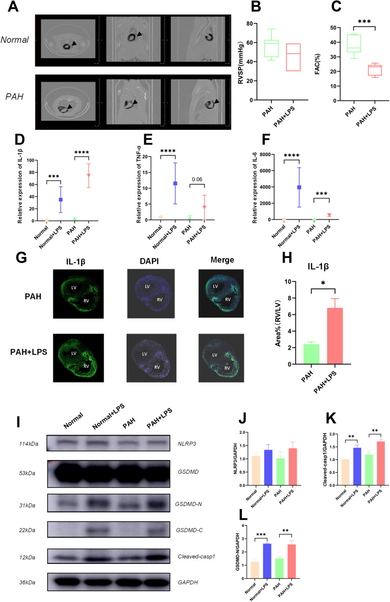
Figure 1.
Activation of the NLRP3 pathway was associated with RVF in PAH rats. (A) PET-CT image of PAH rat hearts showing a thickened wall and dilated chamber of the right ventricle in a rat with pulmonary hypertension. (B) The RVSP of the PAH rats before and after LPS injection is shown. The significance of differences was determined with unpaired Welch’s t-test (n = 6). (C) The FAC [FAC = (end-diastolic area - end-systolic area)/end-diastolic area×100%] in the PAH rats before and after LPS injection is shown. The significance of differences was analysed with unpaired Welch’s t-test (n=6). (D) The IL-1β mRNA level in normal rats (SD rats were fed for 28 days after intraperitoneal injection of saline, and the samples were collected 6 hours after another intraperitoneal injection of saline), normal+LPS rats (SD rats were fed for 28 days after intraperitoneal injection of saline, and the samples were collected 6 hours after intraperitoneal injection of LPS), PAH rats (SD rats were fed for 28 days after intraperitoneal injection of MCT, and the samples were collected 6 hours after intraperitoneal injection of saline), and PAH+LPS rats (SD rats were fed for 28 days after intraperitoneal injection of MCT, and the samples were collected 6 hours after intraperitoneal injection of LPS). The significance of differences among groups was determined by one-way ANOVA (n = 5). (E) The TNF-α mRNA level in the right ventricles of rats in the abovementioned four groups is shown. The significance of differences among groups was determined by one-way ANOVA (n = 5). (F) The IL-6 mRNA level in the right ventricles of rats in the four abovementioned groups is shown. The significance of differences among the groups was determined by one-way ANOVA (n = 5). (G) IL-1β immunofluorescence in the right ventricle of PAH rats and PAH+LPS rats 6 h after LPS injection. (H) LPS significantly increased IL-1β expression in the right ventricle compared with the left ventricle in rats with pulmonary hypertension. The ratio of the IL-1β area% in the right ventricle to that in the left ventricle is shown. The significance of differences was determined with an unpaired Welch’s t-test (n = 3). (I) NLRP3, cleaved-casp1, GSDMD, GSDMD-N and GAPDH protein expression in the right ventricles of rats in the abovementioned four groups. (J) NLRP3 is an important upstream molecule of the pyroptosis pathway, which is involved in the formation of inflammasomes and cleavage of pro-casp1. The NLRP3 protein level in the right ventricle of rats in the abovementioned four groups is shown. The significance of the difference was analysed by one -way ANOVA (n = 3). (K) Cleaved casp1 cleaved the IL-1B precursor to generate mature IL-1B and cleaved GSDMD to expose GSMDM-N, thereby promoting cell lysis and pyroptosis. The cleaved casp1 protein level in the right ventricles of rats in the abovementioned four groups is shown. The significance of the difference was analysed by one -way ANOVA (n = 3). (L) GSDMD is an executor of pyroptosis, and the GSDMD N-terminal domain can assemble membrane pores to induce pyroptosis. The GSDMD-N protein level in the right ventricle of rats in the abovementioned four groups is shown. The significance of the difference was analysed by one -way ANOVA (n = 3). *p < 0.05, **p < 0.01, ***p < 0.001, ****p < 0.0001.

