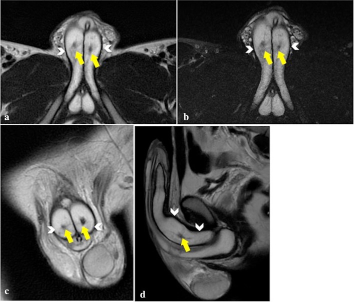
Fig. 2.
MRI axial T2-PROPELLER (a), axial T2 FAT SAT (b), coronal T2-PROPELLER (c), Sagittal T2-PROPELLER (d), images rule out tunica albuginea tears (continuous single hypointense border to the cavernosa—arrowheads) and show low-signal lesions, consistent with arterio-cavernosal fistulas (yellow arrows) not yet visible at first US examination

