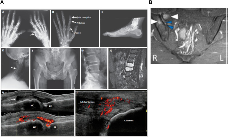
Fig. 4.
Characteristic radiographic features in PsA. Images from A Ritchlin CT, et al. N Engl J Med. 2017;376(10):957–70. 10.1056/NEJMra1505557a and B Braga MV, et al. Sci Rep. 2020;10(1):11580. 10.1038/s41598-020-68456-7b. MC metacarpal head, MRI magnetic resonance imaging, PP proximal phalanx, STIR short-tau inversion recovery. aRadiographic features of PsA: a arthritis mutilans, with pencil-in-cup deformities (arrow) and marked bone resorption (osteolysis) in phalanges of the right hand; b the hand radiograph shows joint resorption, ankylosis, and erosion in a single ray; c enthesophytes at the plantar fascia and Achilles’ tendon insertions; and d syndesmophytes involving the cervical spine, with ankylosis of facet joints (arrow); e bilateral grade 3 sacroiliitis; f paramarginal syndesmophyte bridging the fourth and fifth lumbar vertebrae; g bone marrow edema in the second and third lumbar vertebrae in a patient with severe psoriasis and new onset of back pain; h high-frequency (15 MHz) grayscale ultrasound image shows synovitis of the metacarpophalangeal joint. Distention of the joint capsule is evident (arrows). The confluent red signals (box in the lower part of the image) with power Doppler ultrasonography indicate synovial hyperemia; and i high-frequency (15 MHz) ultrasound image shows enthesitis. The confluent red signals with power Doppler ultrasonography represent hyperemia at the tendon near its insertion into the calcaneus. Normally, the tendon is poorly vascularized [76]. bUnilateral acute sacroiliitis of the sacroiliac joints that can be seen on MRI. Coronal STIR sequence: high signal intensities on the right compatible with bone marrow edema (white arrows) and enthesitis (blue arrows)[75]

