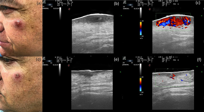
Fig. 1.
Clinical Case 1. Top row: before treatment. a Clinical photograph of a case of cutaneous leishmaniasis with a lesion in the upper cheek. b Grayscale ultrasound: a hypoechoic oval lesion in the dermis with intralesional hyperechoic areas and a lobular hyperechoic involvement of the hypodermis. c Color Doppler: increased intralesional vascularity. Bottom row: after treatment. d Clinical image after 4 weeks of treatment. e Grayscale ultrasound: a decrease in the diameter of the lesion with little involvement of the hypodermis. f Color Doppler: decreased intralesional vascularity

