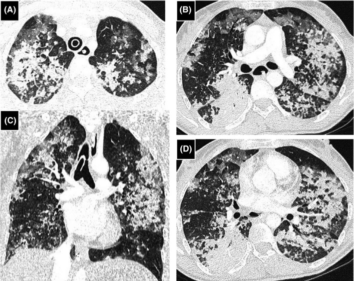
FIGURE 1.

Chest computed tomography (CT) imaging, coronal (C) and axial (A, B, and D) views. Bilaterally multiple patchy ground‐glass opacities, associated with multifocal consolidations and septal thickening

Chest computed tomography (CT) imaging, coronal (C) and axial (A, B, and D) views. Bilaterally multiple patchy ground‐glass opacities, associated with multifocal consolidations and septal thickening