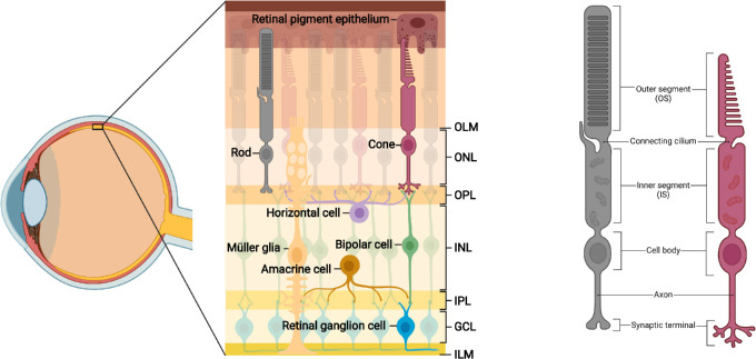
Figure 1.
Organization and circuitry of the retina. (A) The retina contains three layers of cell bodies: the outer nuclear layer (ONL), in which rod and cone cell bodies reside; the inner nuclear layer (INL), containing horizontal cell (HC), bipolar cell (BC), amacrine cell (AC) and Müller glial (MG) cell bodies; and the ganglion cell layer (GCL) where retinal ganglion cell (RGC) somata and displaced ACs are found. PRs are supported by close apposition to the retinal pigment epithelium (RPE). The neural retina is bound apically by the outer limiting membrane (OLM) and basally by the inner limiting membrane (ILM), both formed by end-feet of the MG. PRs connect with BCs and HCs via synapses in the outer plexiform layer (OPL). The inner plexiform layer (IPL) contains signal-carrying synapses between BCs, ACs, and RGCs. (B) Rod and cone PRs display several distinct morphologic features. The outer segment (OS) contains stacked discs of photosensitive opsins for light detection. The connecting cilium facilitates trafficking between outer and inner segments (IS), the latter of which are rich in mitochondria. Extending from the cell body are axons with synaptic terminals, which interact with inner retinal neurons at triad ribbon synapses.

