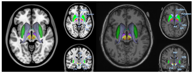
Fig. 1.
Basal ganglia segmentation involved in Parkinson’s disease. Left represents segmentations overlaid on top of MNI template, right represents an example segmentation of a PD subject used in this study (Patient 3127), overlaid on the same subject’s T1w image. Structures segmented are left and right putamen, caudate, nucleus accumbens (NA), globus pallidus externus (GPe), globus pallidus internus (GPi), substantia nigra pars compacta (SNc), substantia nigra pars reticula (SNr), subthalamic nucleus (STN), parabrachial pigmented nucleus (PBP), ventral tegmental area (VTA).

