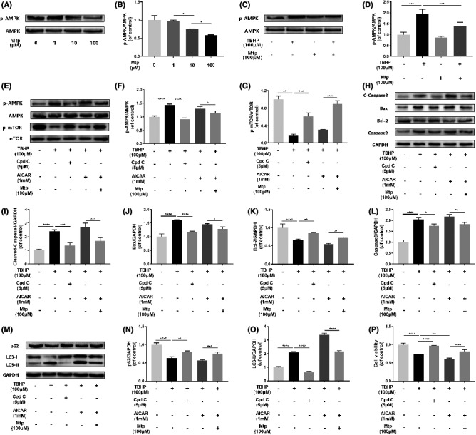
FIGURE 7.

Suppressed of AMPK by Mtp attenuates oxidative stress‐mediated cell apoptosis and autophagy. (A, B) Western blot analysis of expression of p‐AMPK in different dose of Mtp treatment. BM‐EPCs were treated with 1, 10, 100 μM Mtp. The protein expression of p‐AMPK was decreased after Mtp treatment; (C, D) Western blot analysis of expression of p‐AMPK after Mtp pretreatment. Cells pretreated with 100 μM Mtp followed by TBHP stimulation. Mtp‐reduced p‐AMPK protein expression of BM‐EPCs induced by TBHP; (E–O) Western blot analysis of expression of p‐AMPK, p‐mTOR, cleaved‐caspase3, Bax, Bcl‐2, caspase9, SQSTM1/P62 and LC3‐II. Cells were pretreated with 5 µM compound C or 1 mM AICAR for 2 h followed by 100 µM Mtp for 48 h and incubated with TBHP for 3 h. (P) Cell Counting Kit‐8 (CCK‐8) results of BM‐EPCs were treated under the same conditions as above. Cell viability was significantly increased by Cpd C treatment. The densitometric analysis of all Western blot band intensities was normalized to the total proteins or GAPDH. n = 3 independent experiments. *p < 0.05, **p < 0.01, ***p < 0.005 and ****p < 0.001 versus the indicated group
