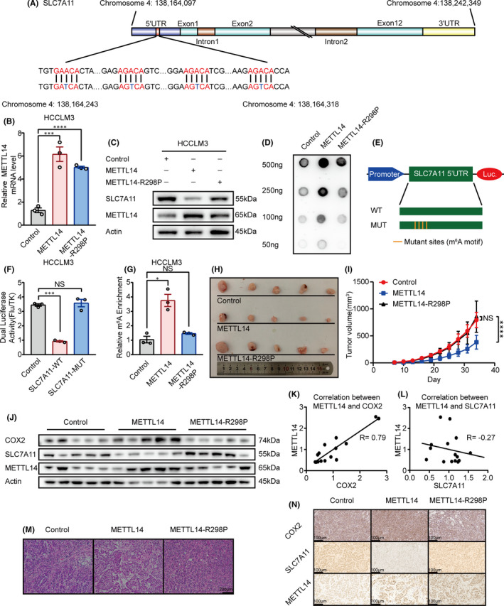
FIGURE 3.

METTL14 triggers m6A methylation of SLC7A11 mRNA at 5’UTR and the anti‐tumour effect of METTL14 is dependent on its methylation activity in HCC. (A) Schematic diagram of SLC7A11 mRNA and the predicted ‘m6A’ sites at 5‘UTR are highlighted in red. Base A in the middle of ‘DRACH’ was replaced by T to make the mutant plasmid for luciferase reporter assay. (B&C) The effect of wide‐type METTL14 and METTL14‐R298P mutant on SLC7A11 expression in HCCLM3 cells. The mRNA and protein level of SLC7A11 was detected by qPCR and Western blot, separately. (D) DOT BLOT showed the total m6A level HCCLM3 that stably expressed wide‐type METTL14 and METTL14‐R298P mutant. (E) Schematic diagram of the luciferase reporter of SLC7A11. (F) Relative activity of the WT or MUT luciferase reporters based on pGL3‐basic plasmid in METTL14 transfected HCCLM3 cells was determined (normalized to vector control groups). (G) MeRIP analysis followed by RT‐qPCR was applied to assess the m6A modification of SLC7A11 in HCCLM3 expressed wide‐type METTL14 or METTL14‐R298P mutant. The enrichment of m6A in each group was calculated by m6A‐IP/input and IgG‐IP/input. (H) The effect of wide‐type METTL14 and METTL14‐R298P mutant on HCC tumour growth. Nude mice were subcutaneously injected with HCCLM3 cells that stably expressed METTL14, METTL14‐R298P or control vector. Tumour growth was calculated twice every week. (I) Tumour growth curve of stable wide‐type METTL14 or METTL14‐R298P mutant overexpressing HCCLM3 cells (or negative control) in the xenograft model was presented. (J) The expression pattern of COX2, SLC7A11 and METTL14 in the xenograft detected by Western blot. (K) The correlation between METTL14 and COX2 in the xenograft. (L) The correlation between METTL14 and SLC7A11 in the xenograft. M. H&E stained section of three kinds of xenografts. (N) The expression pattern of COX2, SLC7A11 and METTL14 in the xenograft detected by immunohistochemistry. ‘NS’, not significant, *p < 0.05, **p < 0.01, ***p < 0.001, ****p < 0.0001
