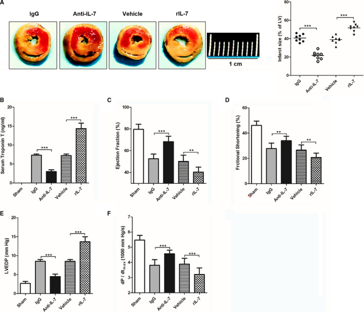
Figure 3.

Effects of IL‐7 neutralization and supplementation on myocardial I/R injury in mice. The heart tissues of mice were harvested and being analysed, and cardiac function and haemodynamic measurements at 24 h after reperfusion; (A) Representative images of left ventricular (LV) slices in sham group, IL‐7 antibody isotype IgG treatment group (IgG), IL‐7 antibody treatment group (Anti‐IL‐7), solvent group (vehicle) and recombinant IL‐7 supplement group (rIL‐7) (left), and the infract size of LV (white) is statistically compared (right); (C‐G) Serum troponin T (B), ejection fraction (C), fraction shortening (D), left ventricular end‐diastolic pressure (LVEDP) (E) and maximum systolic blood pressure (dP/dtmax) (F) in different group mouse at 24 hours after reperfusion. Data shown are mean ± SD (n = 8). Comparison between the two groups, *** was P < .001. Scale bar = 1 cm
