
Figure 4.
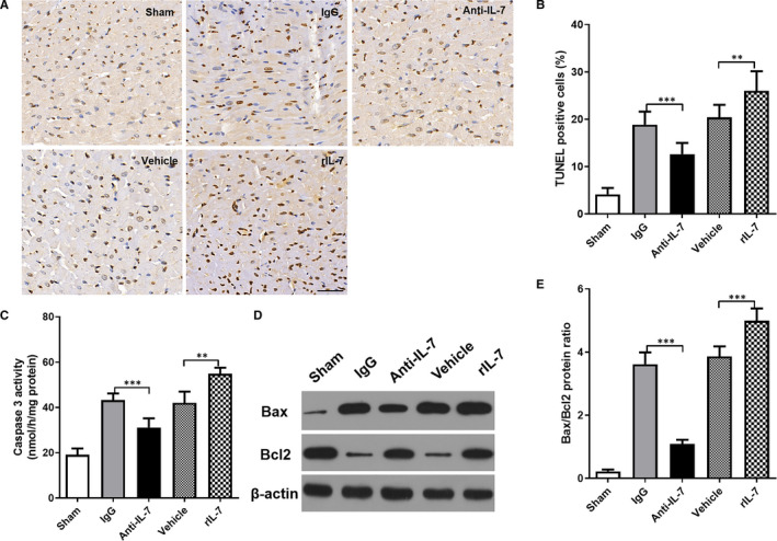
Effects of IL‐7 neutralization and supplementation on cardiomyocyte apoptosis in mice. (A, B) TUNEL staining was used to detect the apoptosis in the heart tissues at 24 hours after reperfusion (A), and statistical comparison of TUNEL positive cells (brown staining) (B); (C) The caspase‐3 activity of the heart was measured and compared; (D, E) Representative images of Bax and Bcl2 protein brands using Western blot in the heart tissues (D), statistical comparison of protein band grey values (E). Data shown are mean ± SD (n = 8). Comparison between the two groups, *** was P < .001