FIGURE 2.

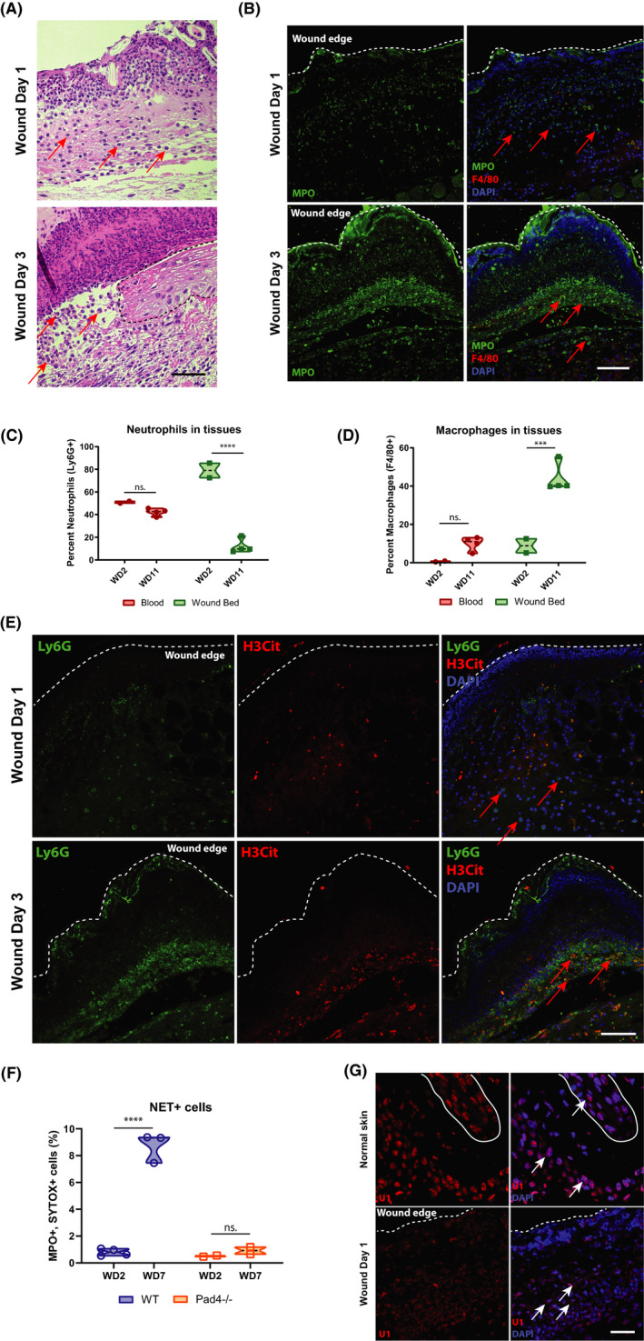
Neutrophils persist in wound bed after the acute inflammatory phase, producing extracellular traps. (A) Neutrophils are present in the wound beds of C57BL/6J mice at early time points, as visible in representative haematoxylin and eosin (H&E) staining. Arrows indicate regions of interest, and dashed line demarcates boundaries. Black scale bar = 50 µm. (B) Neutrophils predominate throughout the wound beds of C57BL/6J mice on wound day (WD) 1 and WD3, visible in prominent myeloperoxidase (MPO) immunofluorescence (IF) staining (green). Few macrophages are present (Red, F4/80). White scale bar = 200 µm. (C) Per cent neutrophil (Ly6G+ cells from total CD45+ cells) levels are consistent in the blood throughout the wound time course but drop in the wound bed at WD11, as measured by FACS. ****p < 0.0001, as calculated by two‐way ANOVA. N = 2 vs. 4. Results are representative of at least two independent experiments. (D) Macrophage (F4/80) levels are largely absent from the blood and low in the wound bed during the early phase of healing but increase dramatically at WD11, as measured by FACS. ***p < 0.004, as calculated by two‐way ANOVA. n.s., not significant. N = 2 vs. 4. Results are representative of at least two independent experiments. (E) Citrullinated histone H3 (H3Cit, red) co‐localized with Ly6G+ neutrophils (green), beginning at WD3 in the wound beds of IF‐stained C57BL/6J mice, indicating the formation of extracellular traps. (F) Neutrophil extracellular trap‐positive cells (MPO+, SYTOX green +) are present at late wound time points, but are absent in the wound beds of PAD4−/− mice, as measured by FACs. ****p < 0.0001, as calculated by two‐way ANOVA. N = 7 vs. 4. Results are representative of at least two independent experiments. (G) Cytoplasmic U1 snRNA is present in the wound bed of C57BL/6J mice, while it localized exclusively in the nuclei of unwounded controls, as visualized by representative FISH. The solid white line delineates a hair follicle. White scale bar = 80 µm
