Fig. 3.
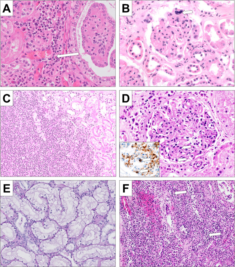
Other morphologic findings in our cohort. A Focus of extramedullary hematopoiesis showing myeloid and erythroid cells as well as a megakaryocyte (arrow) (H&E, × 400). B Extramedullary hematopoiesis showing a megakaryocyte (arrow) in a capillary loop of the glomerulus (H&E, × 400); C Kidney infiltrates of blasts in a patient with transformation of PMF into AML (H&E, × 200); D Glomerulus showing intracapillary blasts and myeloid precursors (arrow) of a case with transformation into AML (H&E, × 400); the insert confirms the blasts in a CD34 immunohistochemical stain (arrow, × 400); E severe acute tubular damage of the osmotic nephrosis type showing fine-vaculoated cytoplasm of the tubuli (H&E, × 200); F acute pyelonephritis showing a dense infiltrate of neutrophils destroying the basement membrane of tubuli (arrow) (H&E, × 200)

