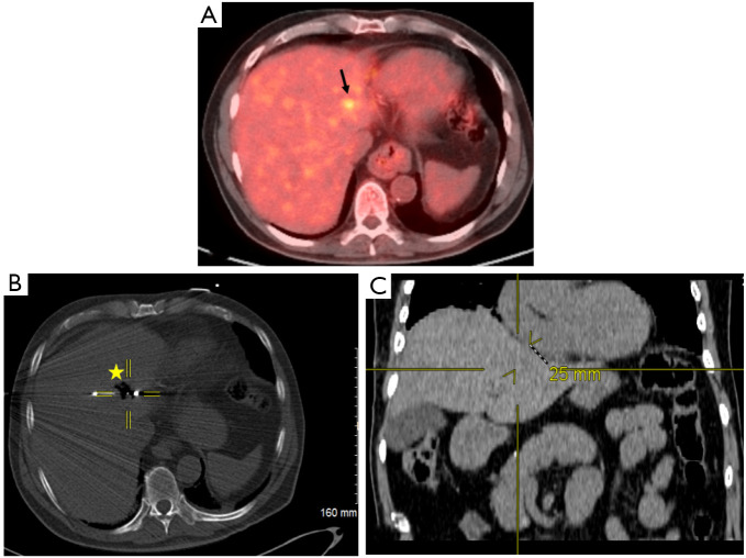
Figure 1.
Localization of primary hepatocellular carcinoma and proximity to the heart. (A) Pre-procedural positron electron tomography/computed tomography (CT) scan demonstrating 11 mm hypermetabolic hepatic lesion (black arrow) in a patient with primary hepatocellular carcinoma. (B) Perioperative CT fluoroscopic imaging demonstrating 4.2 cm spherical zone of ablation (star) with crosshairs centered on the emitter of the microwave antenna. (C) Post-procedural coronal CT image that corresponds to the center of ablation zone. Yellow crosshairs were localized to the same point as center of ablation zone in this coronal view. Distance from center of ablation zone to the heart was measured. It can be noted on the coronal view the center of ablation zone to the heart measures 25 mm in radius corresponding to ≤5 mm distance from the heart.

