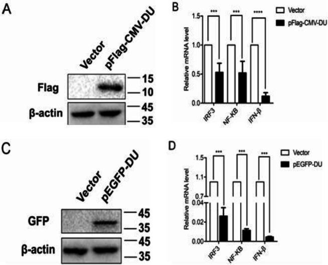
Fig. 1.
Label proteins do not affect the inhibitory effect of DU on IFN-β. (A and C) 293T cells were transfected with pFlag-CMV-DU (0.5 μg) (A), and pEGFP-DU (0.5 μg) (C) and empty vector. The cells were collected at 24 h post-transfection and tested by Western blot. Molecular weight markers in kDa are shown on the right. (B and D) 293T cells were transfected with pFlag-CMV-DU (B), pEGFP-DU (D), and empty vector. Twelve hours after transfection, cells were infected with 0.5 MOI SeV. The infected cells were collected at 24 h post-infection, mRNA levels of IRF3, NF-κB, and IFN-β were tested by real time quantitative reverse transcription-polymerase chain reaction (RT-qPCR). Differences in data considered statistically significant at p-value less than 0.05 (* P<0.05, ** P<0.01, *** P<0.001, and NS: No significant). CMV: Cytomegalovirus, DU: dUTPase, IRF3: Interferon regulatory factor 3, NF: Nuclear factor, IFN-β: Interferon beta, pEGFP: Enhanced green fluorescence protein plasmid, MOI: Multiplicity of infection, and SeV: Sendai virus

