
Figure 1.
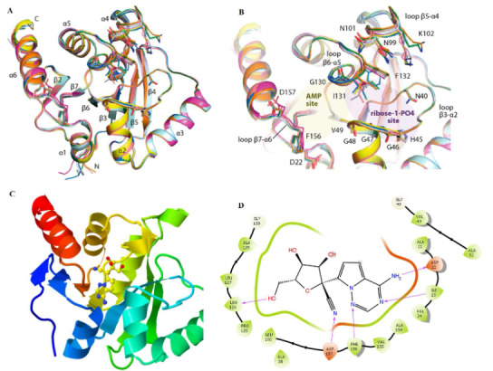
Illustration of the ADP-ribose binding pocket of nsp3 macrodomain of SARS-CoV-2. (A) Plasticity of the ADP-ribose binding pocket; (B) Superimposition of eight molecules from the asymmetric units of two distinct apo crystal structures (PDBs 6YWK and 6YWM) (Reprinted with permission from (Ni et al., 2021), Copyright (2021) American Chemical Society; (C) 3D view of nssp3 Mac-1 in complex with G-441524 (PDB 7BF6); (D) Binding pose showing interactions of G-441524 with amino acids at the active ADP-ribose binding site.