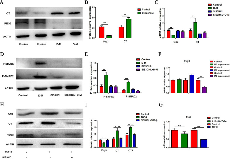
Fig. 10.
Peg3 is involved in the upregulation of OT and OTR expression induced by M2 macrophage polarization. A, B The expression of Peg3 and OT of LMMP in the D-mannose model was detected by western blot analysis. C The levels of Peg3 and OT mRNA of LMMP in the D-mannose model after injection of SIS3HCL (Smad3 inhibitor, 2.5 μg/g) were measured by qRT-PCR. D, E Representative immunoblots for the detection of phosphorylated Smad2/3 after SIS3HCL injection by western blot analysis. β-actin was used to evaluate protein loading. F The level of Peg3 mRNA in cultured enteric neurons treated with or without conditioned medium was measured by qRT-PCR. M2 supernatant suppressed the expression of Peg3. G The level of Peg3 mRNA in cultured enteric neurons treated with or without IL-1β (1 ng/ml), IL-6 (10 ng/ml), TNF-α (20 ng/ml) or TGF-β (10 ng/ml) was measured by qRT-PCR. H, I Cultured enteric neurons were pretreated with SIS3CHL (Smad3 inhibitor, 5 μM) for 12 h followed by TGF-β (10 ng/ml) for 24 h. Only TGF-β inhibited the expression of Peg3. I The protein levels of Peg3, OT, and OTR were analysed by western blot, and β-actin was used to evaluate protein loading. The values represent the mean ± SEM of 6 samples and were compared by t-test or one-way ANOVA with Dunnett’s test for multiple comparisons. *p < 0.05, **p < 0.01, ***p < 0.001

