Fig. 2.
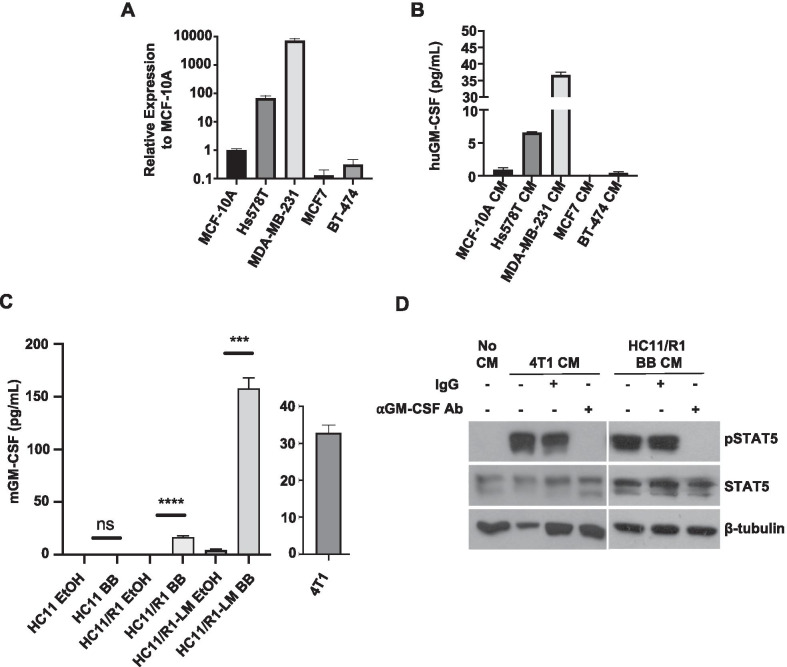
Tumor cell-derived GM-CSF activates STAT5 in macrophages. A qRT-PCR analysis for GM-CSF in TNBC (Hs578T and MDA-MB-231), ER + (MCF7) and HER2 + (BT-474) human breast cancer cells relative to expression in MCF-10A cells. B ELISA analysis for GM-CSF in CM collected from MCF-10A, MDA-MB-231, Hs578T, MCF7, and BT-474 cells. C ELISA analysis for GM-CSF in CM collected from 4T1 cells and B/B-stimulated HC11, HC11/R1, and HC11/R1-LM cells relative to EtOH controls. D Immunoblot analysis for pSTAT5, TSTAT5, and β-tubulin in BMDMs treated with No CM, tumor CM (4T1 or HC11/R1 BB), or tumor CM incubated for 1 h at 37 °C with 2.5 µg/mL neutralizing GM-CSF antibody (⍺GM-CSF Ab)

