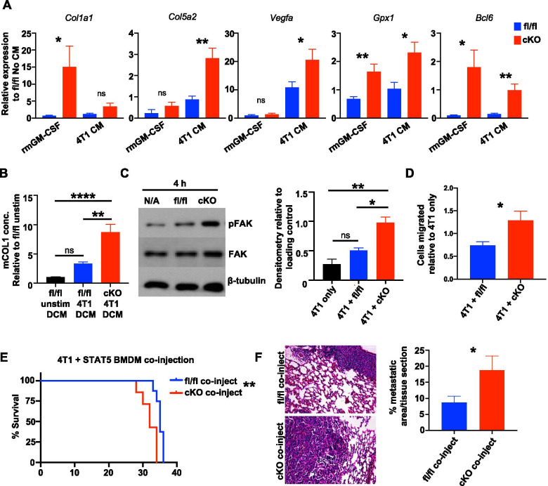
Fig. 5.
STAT5 deletion in macrophages enhances tumor-promoting phenotype and impacts tumor cell migration and metastasis. A qRT-PCR analysis of genes of interest from RNA-seq associated with tumor-promoting pathways in rmGM-CSF or 4T1 CM-treated STAT5fl/fl (blue) and STAT5cKO (red) BMDMs. Unpaired t-test was used for statistical analysis. B Mouse Type 1 Collagen ELISA in STAT5fl/fl unstimulated or 4T1 CM-stimulated STAT5fl/fl and STAT5cKO macrophage double CM (DCM). Data were analyzed using one-way ANOVA and Tukey’s multiple comparison test. C Representative immunoblot of pFAK, total FAK (FAK), and β-tubulin in 4T1 cells cultured alone or co-cultured with STAT5fl/fl or STAT5cKO BMDMs for 4 h. Densitometry analysis relative to loading control. D Migration analysis of 4T1 cells cultured alone or co-cultured with STAT5fl/fl or STAT5cKO BMDMs after 20 h. Cell counts relative to 4T1 alone in triplicate. E Kaplan–Meier curves of 4T1 cells co-injected with either STAT5fl/fl (n = 8) or STAT5cKO (n = 7) BMDMs in WT BALB/c mice. % Survival on Y-axes indicates proportion of mice reaching tumor size endpoint of 1cm3. F Quantified metastasis in H&E-stained lung sections. Lungs were sectioned at 3 different depths per mouse and analyzed for percent metastatic area per tissue section. *P < 0.05, **P < 0.01, ***P < 0.001, ****P < 0.0001. Scale bar: 50 μm

