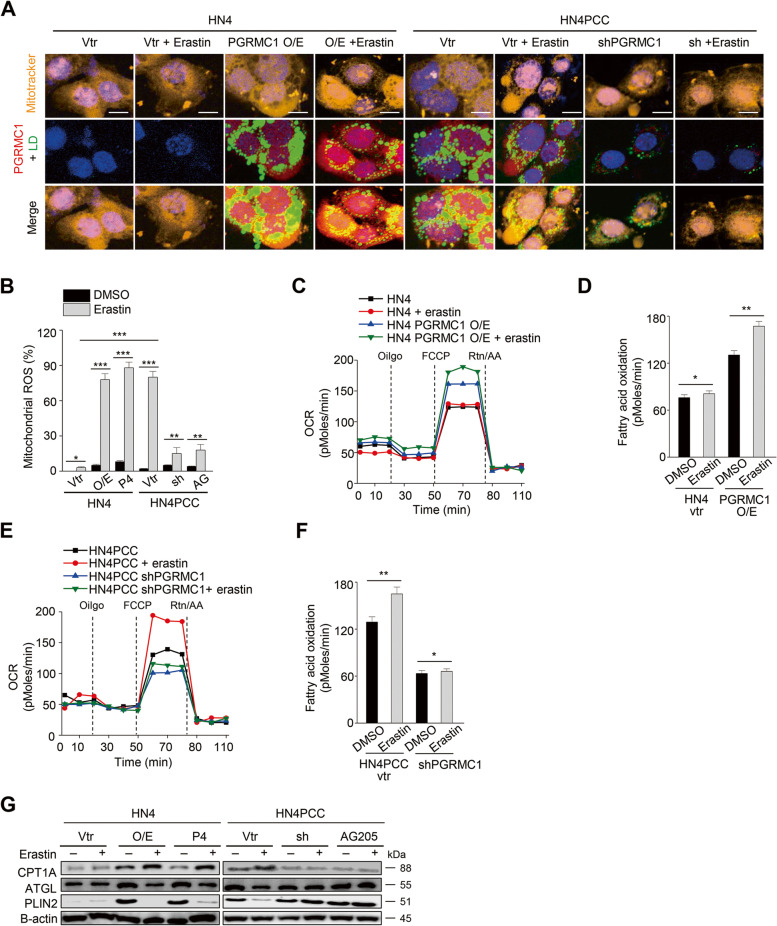
Fig. 6.
PGRMC1 increases FAO by anchoring lipid droplets to mitochondria. A Co-staining of mitotracker (orange), lipid droplets (LD, green), and PGRMC1 (red) in HN4 parental cells and PCC with or without 10 μM erastin treatment for 24 h. HN4 cells were transfected with vector (vtr) or PGRMC1 overexpression vector or treated with 100 nM P4. HN4PCC were transfected with vector or shPGRMC1 or treated with 20 μM AG205. Scale bar 5 μm. B Mitochondrial ROS was examined using incubation with 5 μM mitoSOX™ Red and FACS in HN4 parental cells and PCC with or without PGRMC1 overexpression or inhibition and with DMSO or 10 μM erastin for 8 h. Data are means and s.d. from three technical replicates. *P < 0.05, **P < 0.01, ***P < 0.001 relative to DMSO control or between different groups. C-F Quantification of FAO in HN4 parental cells and PCC with or without PGRMC1 overexpression or inhibition and with DMSO or 10 μM erastin. *P < 0.05, **P < 0.01 relative to DMSO control. G Immunoblotting in HN4 parental cells and PCC with or without PGRMC1 overexpression or inhibition and with DMSO or 10 μM erastin for 24 h. O/E, PGRMC1 overexpression vector; sh, shPGRMC1; P4, progesterone; AG, AG205

