. 2021 Aug 24;301(2):295–308. doi: 10.1148/radiol.2021203645

Figure 5:

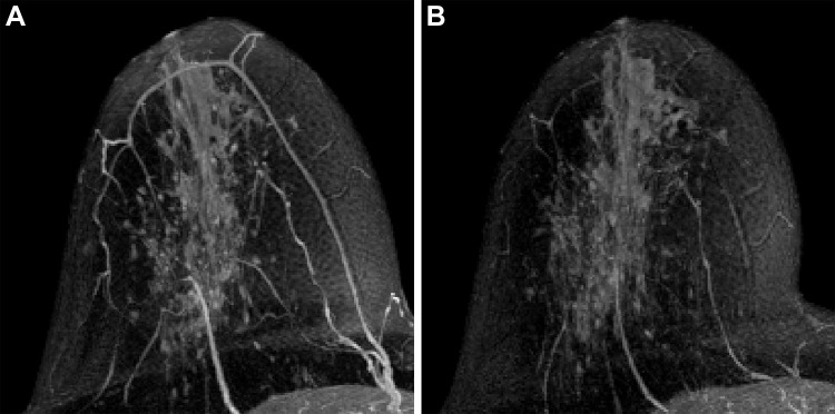
Axial maximum intensity projection of subtracted early contrast-enhanced phase MRI in right breast of a 55-year-old postmenopausal woman with left hormone receptor–positive human epidermal growth factor 2–negative invasive breast cancer at (A) T0 and (B) T2. The calculated background parenchymal enhancement (BPE) values were (A) 32.3% and (B) 35.0%, and the BPE change was evaluated as nonsuppressed (percent change of BPE at T2, ≥0). The patient was confirmed at pathologic analysis as having noncomplete response in the surgical specimen after neoadjuvant chemotherapy.

Axial maximum intensity projection of subtracted early contrast-enhanced phase MRI in right breast of a 55-year-old postmenopausal woman with left hormone receptor–positive human epidermal growth factor 2–negative invasive breast cancer at (A) T0 and (B) T2. The calculated background parenchymal enhancement (BPE) values were (A) 32.3% and (B) 35.0%, and the BPE change was evaluated as nonsuppressed (percent change of BPE at T2, ≥0). The patient was confirmed at pathologic analysis as having noncomplete response in the surgical specimen after neoadjuvant chemotherapy.