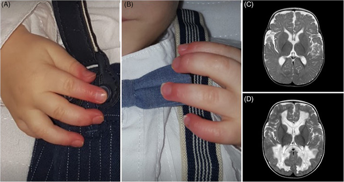
FIGURE 1.

Finger findings and cerebral MRI in the reported patient. A and B, Chronic distal phalangeal erythema, an accompanying sign since the age of 5 months old. C, Axial T2W at the age of 12 months old showing mild cerebral atrophy. D, Axial T2W at the age of 24 months old with appearance of cystic change in white matter. White matter tracts surrounding the lateral ventricles and commissural fibres had signal abnormalities, showing small necrotic or porencephalic cysts. “U” fibres, pyramidal tracts and gray matter were spared
