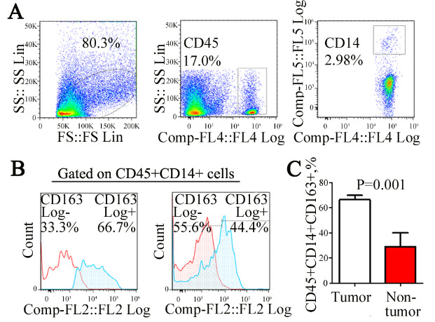
Figure 1.

Expression ratio of CD163+ mono-macrophage in bladder cancer tissue. Mono-macrophages infiltrating the tissue were detected by flow cytometry (A). Notably, 80.3% of living cell enrichment regions in total single cell suspension were obtained from tumor tissue homogenate. Approximately, 17.5% mononuclear macrophages were detected in 80.3% of leukocytes in the preceding gate. In total, 2.98% mononuclear macrophages were detected in 17.5% of leukocytes in the preceding gate. The expression of the CD45+CD14+CD163+ mono-macrophage subset in bladder cancer tissues (n = 8) was significantly higher than that in adjacent normal tissues (B and C) (n = 3, 67.88 ± 7.2% vs. 27.05 ± 11.18%, p = 0.001).
