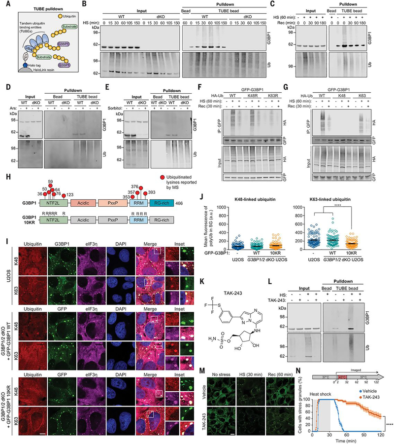
Fig. 1. G3BP1 undergoes K63-linked ubiquitination in response to heat stress.
(A) Illustration showing TUBE capture of ubiquitinated G3BP1. TEV, tobacco etch virus protease cleavage site. (B and C) Immunoblots of TUBE-captured cell extracts showing levels of ubiquitinated G3BP1 in U2OS cells after different durations of 43°C heat shock, using U2OS G3BP1/2 dKO cells as controls (B), and during 37°C recovery (C). (D and E) Immunoblots of TUBE-captured cell extracts showing levels of ubiquitinated G3BP1 in response to oxidative stress (0.5 mM NaAsO2, 1 hour) (D) or osmotic stress (0.4 M sorbitol, 1 hour) (E). (F and G) Immunoblots of cell extracts captured with antibody to GFP, showing K63-linked ubiquitination of G3BP1. Transfected HEK293T cells were exposed to heat shock (43°C, 1 hour) and recovery (37°C, 30 min). K48R and K63R prevent the formation of K48-linked (K48R) or K63-linked (K63R) chains; K48 and K63 permit K48-linked or K63-linked chains exclusively. HA, hemagglutinin; IP, immunoprecipitate. (H) G3BP1 domain labeled with lysines on which ubiquitination has been reported. In the G3BP1 10KR mutant, six lysines in NTF2L and four lysines in RRM are mutated to arginine. (I) Immunofluorescent staining of fixed U2OS WT and U2OS G3BP1/2 dKO cells stably expressing GFP-G3BP WT and 10KR. Scale bar, 10 μm. (J) Fluorescence intensities of K48- and K63-linked ubiquitin in eIF3η-positive stress granules from three technical replicates are plotted in (n = 90). Error bars indicate SEM. ****P < 0.0001 (ANOVA with Tukey’s test). (K) Structure of TAK-243. (L) Immunoblot of TUBE-captured cell extracts showing block of heat shock–induced G3BP1 ubiquitination by TAK-243. U2OS cells were treated with DMSO or TAK-243 for 1 hour prior to heat shock. (M and N) Fluorescent imaging of U2OS cells stably expressing GFP-G3BP1 were treated with DMSO or TAK-243 (1 hour) prior to imaging. Representative images are shown in (M). In (N), GFP signals were monitored at 30-s intervals to count cells with two or more stress granules from three technical replicates (vehicle n = 32, TAK-243 n = 35). Scale bar, 20 μm. Error bars indicate SEM. ****P < 0.0001 (Mantel-Cox test).

